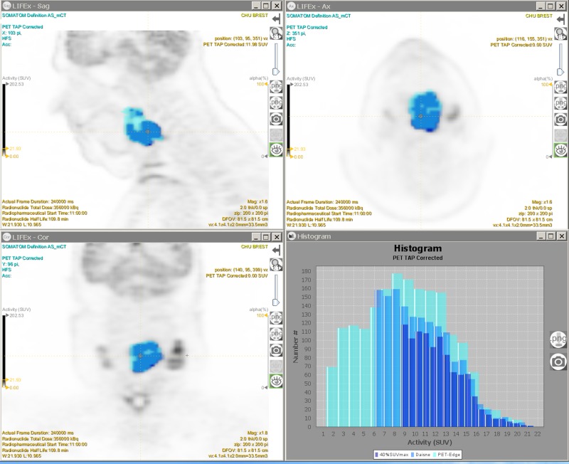
Fig 4. Example of VOI delineating a tumor with the 3 segmentation methods.
(Turquoise blue) PET-EDGE segmentation method. (Sky blue) DAISNE segmentation method. (Dark blue) 40%SUVmax segmentation method. (Top left) FDG PET sagittal slice. (Top right) FDG PET transverse slice. (Bottom left) FDG PET frontal slice. (Bottom right) SUV histograms. With LIFEx software.

