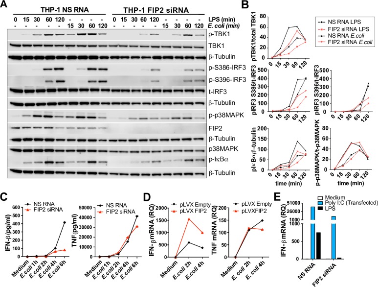
Fig 7. FIP2 controls E. coli induced IFN-β mRNA induction and secretion.
(A) Immunoblots showing the phosphorylation patterns of TBK1, IRF3, IκBα and p38MAPK in FIP2 silenced THP-1 cells stimulated with E. coli bioparticles or LPS (100 ng/ml). Data are representative of three independent experiments. (B) Quantification of phosphorylation patterns of the proteins shown in the immunoblots presented in (A). (C) ELISA quantification IFN-β and TNF secretion in THP-1 cells treated with NS RNA or FIP2 siRNA and stimulated as indicated. (D) Quantification of E. coli-stimulated IFN-β and TNF mRNAs in THP-1 cells with lentiviral overexpression of FIP2. (E) Quantification of Poly I:C and LPS stimulated IFN-β mRNA induction in cells treated with NS RNA or FIP2 siRNA after 4 hours of stimulation. Poly I:C (5 μg/ml) was transfected using Lipofectamine® 2000. Data are representative of three independent experiments.

