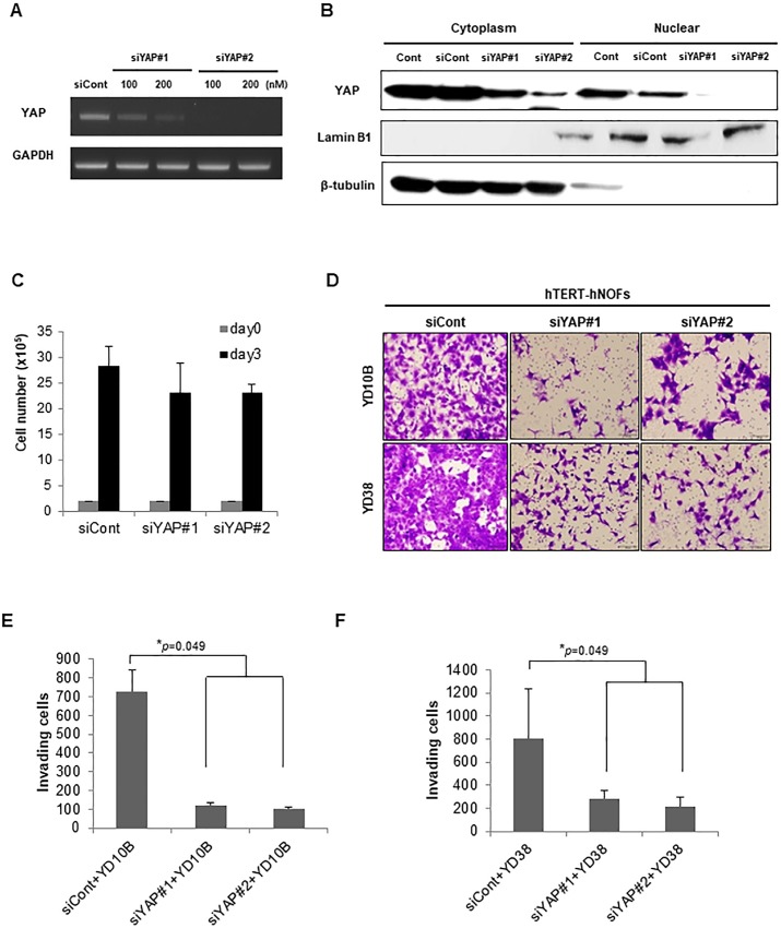
Fig 6. YAP-silencing fibroblasts attenuate invasiveness in OSCC cells.
(A) The depletion of YAP with 2 different siRNAs was identified by RT-PCR. Representative image is shown. (B) Nuclear/cytoplasmic fraction was isolated from each protein and then western blot was performed to check nuclear/cytoplasmic YAP expression by siRNAs targeting YAP. Lamin B1 was used as a positive control of nuclear protein fraction and β-tubulin was used as a positive control of cytoplasmic protein fraction. (C) Cell proliferation in siCont- and siYAPs-transfected hTERT-hNOFs. Cells (2 x 10⁵) were seeded into well and then counted after 3 days. (D) Representative images of invading OSCC cells toward lower chamber are shown (magnification: 200X, scale bar: 100 μm). (E, F) The bar graphs indicated a quantification of invading YD10B (E) or YD38 (F) OSCC cells as siYAPs-transfected hTERT-hNOFs were placed in the lower chamber by comparison with siCont transfected hTERT-hNOFs. This assay (E and F) was performed in independent triplicate repeats (*p < 0.05; Mann-Whitney U test).

