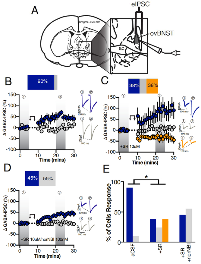
Figure 1. Endogenous neuropeptides modulation of electrically-evoked ovBNST GABAA-IPSCs.
A, Schematic illustrating stimulating and recording electrodes placement in mice brain slices containing the ovBNST. Recordings were restricted to the displayed shaded oval area. B-D, Effects of postsynaptic depolarization (double arrow symbol) on binned (1 minute, 6 events) electrically-evoked GABAA-IPSCs in (B) aCSF (n=10 cells/6 mice), (C) the presence of the non-selective NTR antagonist SR142948 (10μM, n=8 cells/5 mice) or (D) SR142948 + the KOR antagonist norNBI (100nM, n=11 cells/4 mice). Insets in B-D are representative electrically-evoked GABAA-IPSCs before and after postsynaptic depolarization (double arrows). E, Bar graphs summarizing the proportion of responding neurons to postsynaptic depolarization across different pharmacological treatments. Blue LTP, grey no change and orange LTD. Asterisks, p<0.05.

