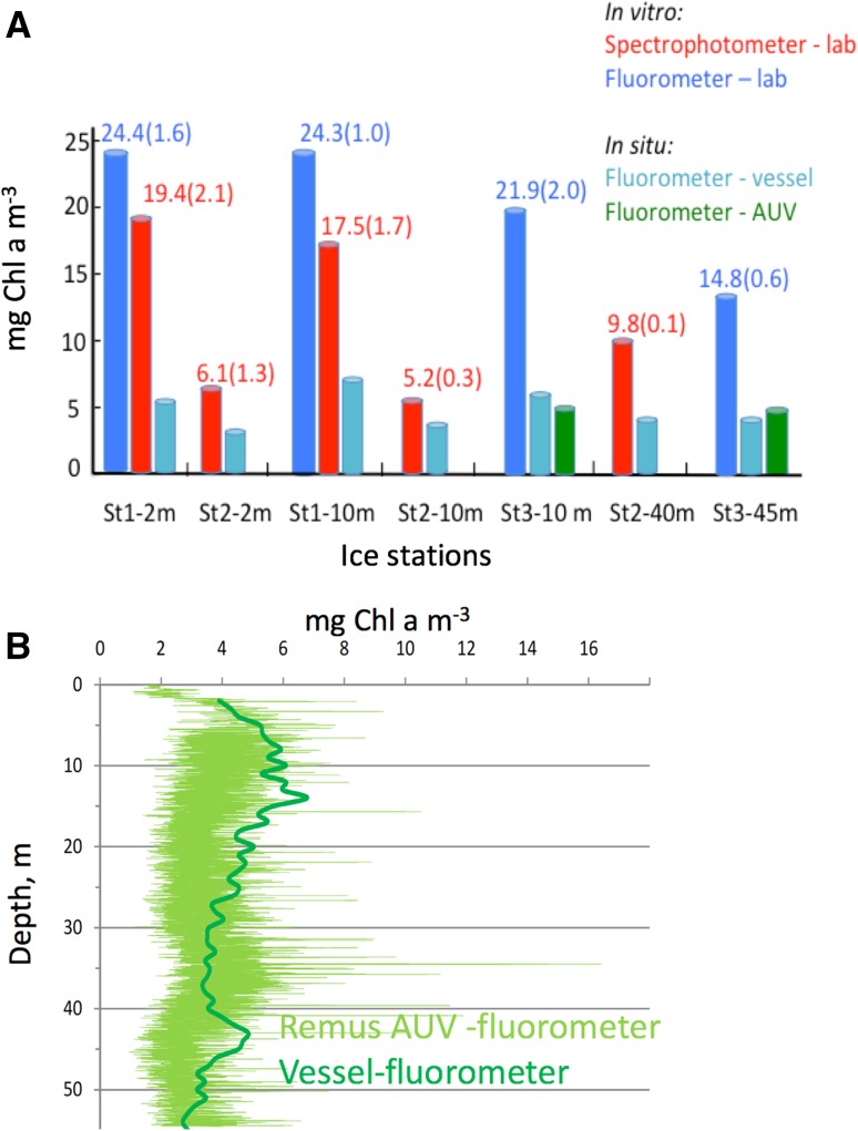
Fig. 7.
a Differences in in situ (living cells) versus in vitro (pigment extracts) [Chl a] at ice station (St) 1–3. These differences may mainly be due to quenching of Chl a fluorescence due to PQ and NPQ in living cells. Numbers in parentheses indicate ± SE, n = 3 for lab-based in vitro measurements. b In situ [Chl a] from repetitive AUV vertical zig-zag transects at ice station 3 (see also Fig. 5) compared to corresponding ship based vertical [Chl a] profile (mean of downcast and upcast in nearby ice-lead)

