. 2019 Mar 28;8:e39964. doi: 10.7554/eLife.39964

Figure 2. Overexpression of Patronin causes dendrite pruning defects in ddaC neurons.

(A–E) Live confocal images of ddaC neurons expressing mCD8-GFP driven by ppk-Gal4, Gal44-77 or ppk-CD4-tdGFP at WP and 16 hr APF. While ddaC neurons overexpressing the UAS control construct pruned all the dendrites (A), ddaC neurons overexpressing Patronin (B) driven by Gal44-77 or GFP-Patronin (C) driven by two copies of ppk-Gal4 at a higher level exhibited simple arbors at WP stage and consistent dendrite pruning defects at 16 hr APF. ddaC neurons, in which GFP-Patronin was expressed by GeneSwitch-Gal4-2295 in RU486-induced conditions (E), exhibited normal arbors at WP stage but severe dendrite pruning defects at 16 hr APF, compared to those in non-induced conditions (D). Red arrowheads point to the ddaC somas. (F) Quantification of total length of unpruned ddaC dendrites at 16 hr APF. (G) Quantification of severing defects at 16 hr APF. Scale bar in (A) represents 50 μm. Error bars represent SEM. The number of samples (n) in each group is shown on the bars. ***p<0.001 as assessed by two-tailed Student’s T test.

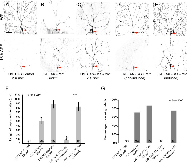
Figure 2—source data 1. Extended statistical data as Microsoft Excel spreadsheet.
DOI: 10.7554/eLife.39964.009

Figure 2.

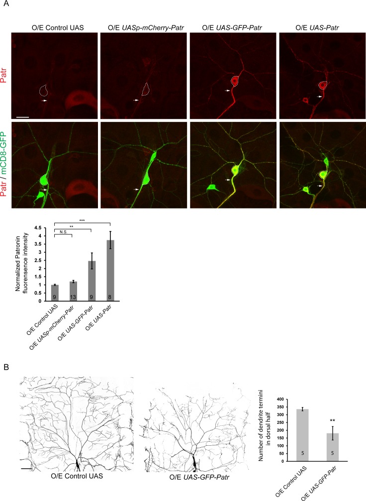
Figure 2—figure supplement 1. Overexpression of Patronin causes dendrite arborization defects in ddaC neurons.

Figure 2—figure supplement 1.

(A) Confocal images of ddaC neurons expressing mCD8-GFP and immunostained for anti-Patronin at w3L stage. ddaC somas are marked by dashed lines, and axons by arrows. Endogenous Patronin was detected in the ddaC neurons expressing an unrelated UAS control. Exogenous Patronin was expressed at a low level in ddaC neurons using a UASp-mCherry-Patronin transgene, but at much higher levels using UAS-GFP-Patronin or UAS-Patronin transgenes. Quantification of normalized Patronin fluorescence intensity in the somas of ddaC neurons. (B) Live confocal images of ddaC neurons expressing mCD8-GFP driven by two copies of ppk-Gal4 at w3L stage. Compared to the control neurons, GFP-Patronin-overexpressing ddaC neurons showed simple dendrite arbors at wL3 stage. Quantification of number of dendrite termini in the dorsal half of ddaC neurons. Error bars represent SEM. The number of neurons (n) in each group is shown on the bars. Scale bars in (A) and (B) represent 20 μm and 50 μm, respectively. N.S., not significant; **p<0.01; ***p<0.001 as assessed by one-way ANOVA test or two-tailed Student’s T test.
Figure 2—figure supplement 1—source data 1. Extended statistical data as Microsoft Excel spreadsheet.
DOI: 10.7554/eLife.39964.008