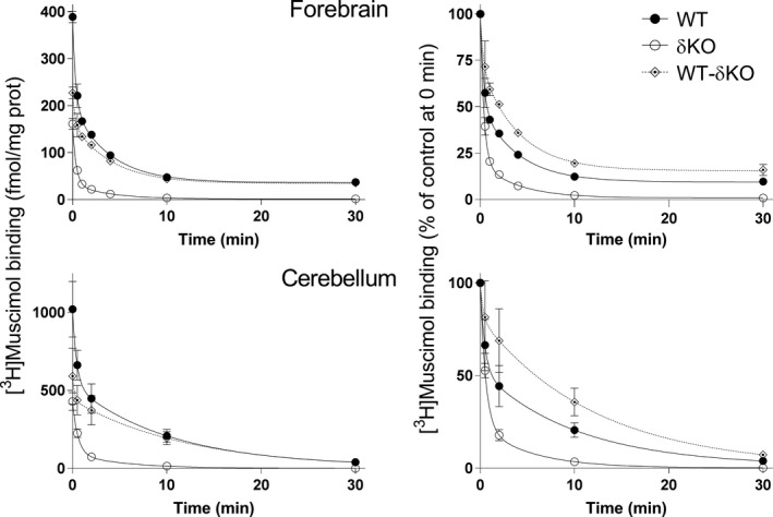
Figure 3.

The δ subunit leads to very slow muscimol dissociation. Dissociation of 5 nM [3H]muscimol binding from forebrain (n = 4 independent experiments using individual forebrains in each experiment) and cerebellar (n = 3 independent experiments using pools of 3 individual cerebella from the mouse line in each pool) membranes of WT and δKO mice (mean ± SEM). The experiments were performed in triplicate technical replicates. Forebrain and cerebellar membranes of the mouse lines were pre‐incubated for 15 min with 5 nM [3H]muscimol alone and in the presence of 100 μM GABA to determine non‐specific binding. Then 100 μM GABA was added to all tubes to start [3H]muscimol dissociation. The incubations were continued for various durations (30 s to 30 min) and terminated by filtration onto GF/B filters. The values are expressed as fmol/mg protein on the left and as % of control binding at the start of dissociation (0 min) on the right. The values for δ‐GABAARs (WT‐δKO) were calculated as described in Materials and methods.
