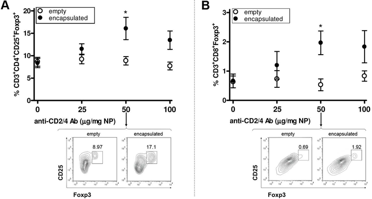
Figure 2. Establishment of the conditions for in vitro induction of CD4 and CD8 Tregs with anti-CD2/CD4 Ab-coated PLGA NPs encapsulated with IL-2 and TGF-β.
Sorted CD3+ T cells (negatively selected with magnetic beads) from 12 weeks-old BALB/c mouse splenocytes were cultured in the presence of anti-CD3/28 Ab and scalar doses of NPs. After three days, flow cytometry analysis compared the numbers of CD4+ Tregs (A) and CD8+ Tregs (B) in cultures with NPs encapsulated with IL-2/TGF-β (closed circles) vs. cultures with empty NPs (empty circles). Significant differences were observed at a dose of 50 μg/mg NP (*P<0.05), for which the figure includes representative flow cytometry plots on pregated CD4 (A) and CD8 (B) cells. Representative of four experiments, each with three mice per group.

