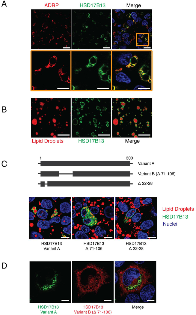
Figure 3.
HSD17B13 localizes with lipid droplets. (A) HepG2 cells were transfected with HSD17B13(A)-GFP and stained for the lipid droplet marker protein ADRP (Perilipin-2). (B) Lipid droplets were isolated from HSD17B13(A)-GFP transfected cells and stained with the lipid droplet specific dye LipidTox. (C) Mutations were generated and tested for cellular localization with LipidTox used to stain lipid droplets. (D) HSD17B13(A)-GFP and HSD17B13(B)-Flag were co-transfected into HEK293 cells. Immunofluorescence staining was used to visualize HSD17B13(B)-Flag. Nuclei were counterstained with Hoechst 33342. One representative image from repeated experiments is shown. Bars indicate 10 μm.

