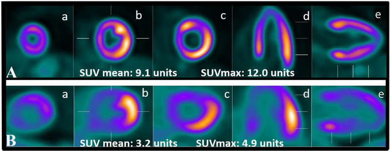
Figure 1: Examples of diffuse and focal myocardial 18F-FDG uptake in two patients with rheumatoid arthritis.
Examples of diffuse (panel A) and focal (panel B) FDG uptake in two different patients with RA. Short axis is represented by a, b and c; horizontal axis in d; and, vertical long axis in e and f.

