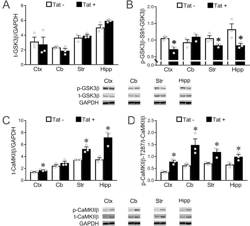
Figure 2. In vivo expression of Tat activates GSK3β and CaMKIIβ in multiple brain regions.
(A) In vivo expression of Tat has no effect on total GSK3β level in any of the four brain regions we examined. (B) In vivo Tat expression leads to upregulated activity of GSK3β (decreased ratio of p-GSK3β-S9/t-GSK3β) in cortex, striatum and hippocampus, but not cerebellum (p = 0.077). (C-D) Western blots show that Tat expression in vivo leads to an increased total CaMKIIβ level in cortex, striatum, and hippocampus (C, cerebellum: p = 0.09), and upregulated CaMKIIβ activity (increased p-CaMKIIβ-T287/t-CaMKIIβ ratio) in all four regions examined (* p < 0.05 vs. corresponding control). In A-D, Ctx: cortex; Cb: cerebellum; Str: striatum; Hipp: hippocampus. Data in A-D are based on the results from N=3 individual Tat+ and Tat− mice, each from a different litter.

