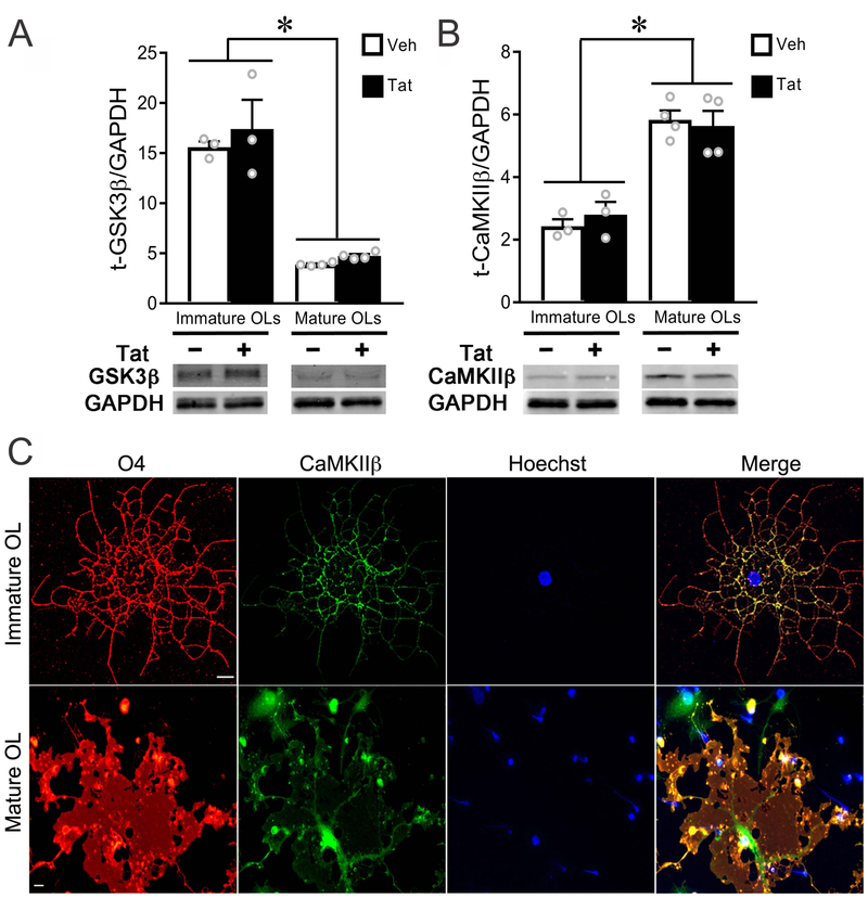
Figure 3. Expression level and localization of CaMKIIβ differ between immature and mature OLs.
(A) Western blotting showed that GSK3β levels were significantly (~3 fold) higher in immature versus mature OLs in vitro, and unaffected by Tat at either stage. (B) Expression of CaMKIIβ was significantly lower in immature versus mature OLs in vitro, and not affected by 96 h Tat treatment. (C) Confocal images (maximum projection) of fluorescent immunostaining showed that CaMKIIβ was expressed in OLs in vitro. In immature OLs, the majority of CaMKIIβ was found in cellular processes. In mature OLs, CaMKIIβ was found both in cell body cytoplasm and along larger processes. (Scale bar: 10 μm; *: p < 0.05, vs. corresponding control). Experiments in A and B represent results from N=3 (immature) or N=4 (mature) independent cultures prepared from mice of different litters.

