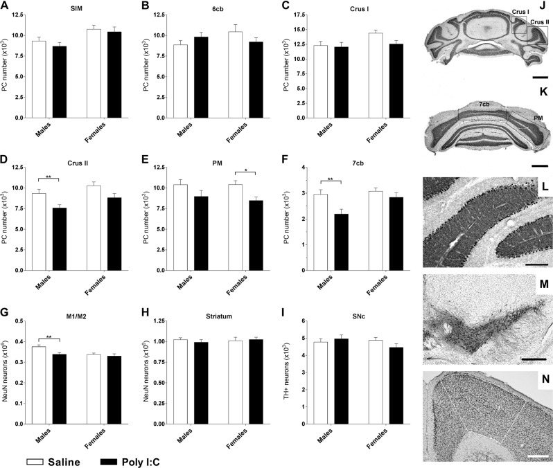
Fig. 4. Prenatal exposure to poly I:C leads to reduced numbers of neurons in the cerebellum and motor cortex in a sex-dependent manner.
a–f Stereological Purkinje cells (PC) counts on coronal sections of the cerebellum. j, k Photomicrographs of the different sub-lobules of lobule VII, scale bars = 1 mm. l Illustration of the monolayer organization of PC in the cerebellar cortex after DAB-calbindin immunolabeling, scale bars = 200 µm. No decrease of the PC number occurred within lobule VI, neither in the hemispheric part, SIM (a) nor in the vermal part, 6cb (b). In the hemispheric part of the lobule VII, the number of PC in Crus I (c) was not affected by treatment whereas a significant reduction was found in Crus II in poly I:C males (d) and in paramedian lobule (PM) in females (e). In the vermal part corresponding to the sub-lobule 7cb (f), the number of PC was significantly decreased in poly I:C males. g Decreased number of NeuN-stained neurons in M1/M2 motor cortex in poly I:C males (outlined area on (n), scale bars = 400 µm). h Unaffected numbers of NeuN-stained striatal neurons or (i) tyrosine hydroxylase-positive neurons in the Substantia Nigra pars compacta (SNc), (m) DAB-TH immunolabelling, scale bars = 400 µm). n = saline males/poly I:C males/saline females/poly I:C females; n (a) = 11/10/10/9; n (b) = 14/11/10/8; n (c) = 13/11/9/9; n (d) = 13/10/9/8; n (e) = 12/8/9/8; n (f) = 12/10/8/8; n (g) = 12/9/10/9; n (h) = 12/9/10/9; n (i) = 12/9/11/9. Data expressed as mean ± SEM; two-way ANOVA followed by Fisher’s LSD or Kruskal-Wallis test followed by Dunn’s multiple comparisons test (b) (*p < 0.05; **p < 0.01)

