Fig. 1.
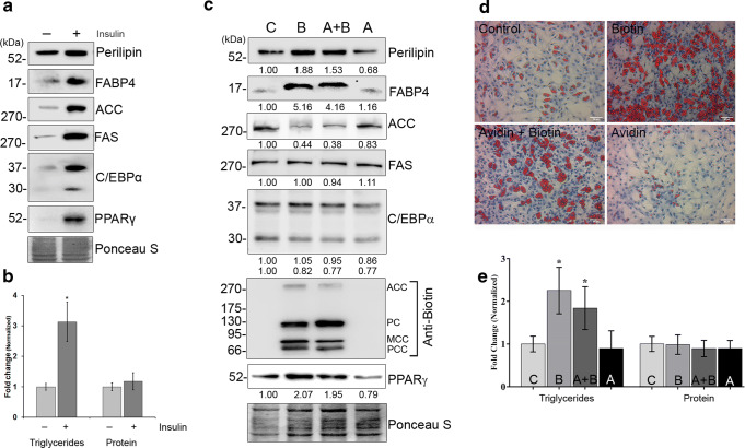
Biotin accelerates insulin induced adipogenesis. [a] Differentiation potential under basal and insulin-stimulated conditions of cultured primary cells was determined using immunoblot by targeting the markers of adipocyte differentiation. [b] Accumulation of triglycerides during adipocyte differentiation. The values were normalized against cellular DNA content and those with p < 0.05 were considered significant. [c] Effects of biotin on lipogenesis were determined by immunoblotting against the markers of adipocyte differentiation. Total proteins were stained using Ponceau S to normalize protein loading. The values were denoted as fold changes of three independent experiments. [d] The phase contrast microscopic images of ρ-formaldehyde fixed cells were stained with oil red O (red) and hematoxylin (blue) to detect lipid droplets and nucleus, respectively (bar 50 μm). [e] Total cell lysates from day 6 of adipocyte differentiation were used to determine the cellular triglyceride and protein content. The values were normalized against their respective DNA content to minimize the variations in cell lysis or loading and represented as fold changes with respect to control (n = 5). *p < 0.05 versus control groups

