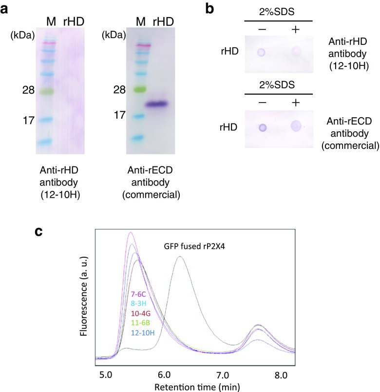
Fig. 1.
Screening of monoclonal antibodies. a Western blotting for rHD using anti-rHD antibody 12-10H (left) and anti-rECD antibody (right). b Dot blot of rHD in the presence (right) or absence (left) of 2% SDS using anti-rHD antibody 12-10H (upper) and anti-rECD antibody (lower). c GFP-fused rat P2X4 in the absence (black) or the presence of 7-6C (magenta), 8-3H (cyan), 10-4G (brown), 11-6B (green), and 12-10H (indigo) was monitored by fluorescence-detection size-exclusion chromatography (FSEC). FSEC was performed with the Superdex 200 5/150 GL at flow rate 0.5 ml/min. The fluorescence was detected at 525 nm with excitation at 490 nm

