Fig. 3.
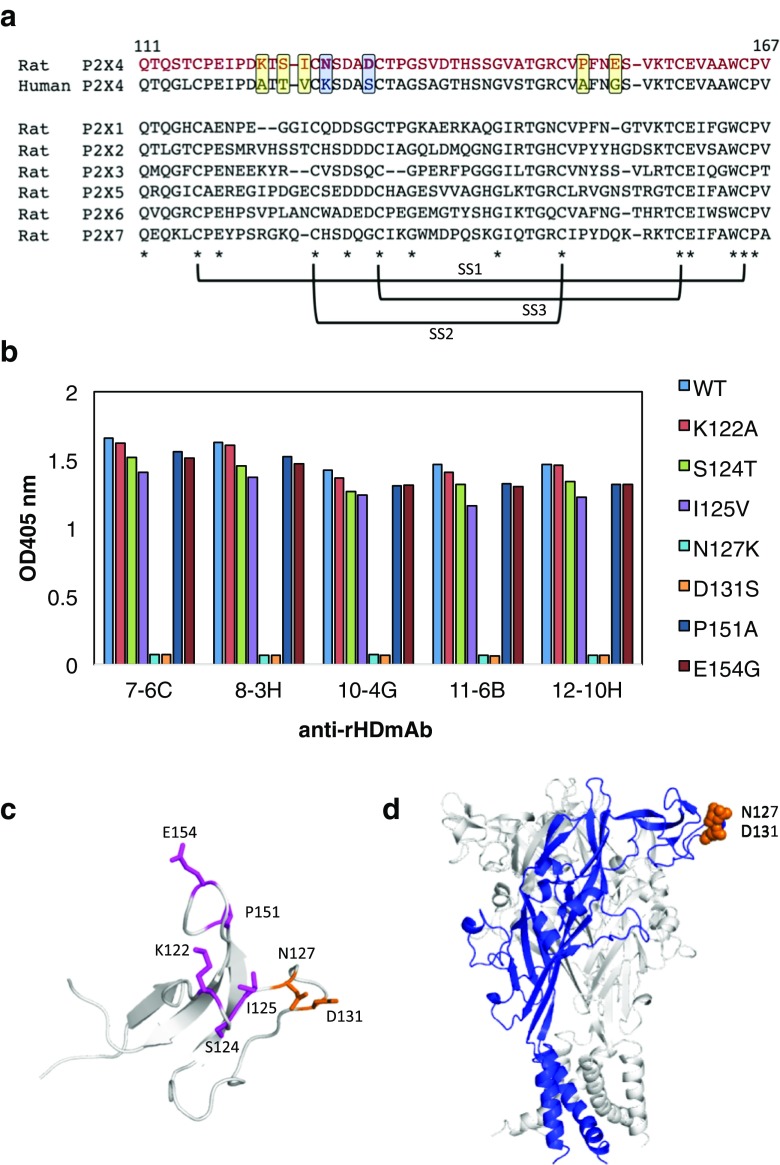
Epitope of rP2X4 for anti-rHD antibody. a Sequence alignment of the head domain of human P2X4 and rat P2X1~P2X7. The mutation sites were enclosed in squares. Conserved amino acids are indicated by asterisks and three S-S bond formations are represented. b The results of ELISA for rHD mutants (K122A, S124T, I125V, N127K, D131S, P151A, and E154G) using each monoclonal antibody as a primary antibody and alkaline phosphatase–conjugated anti-mouse IgG as a secondary antibody. c Mutation sites (stick model) were mapped on the structure of rHD. The epitope is shown in blue. d The epitope is indicated on the homology model of the rP2X4 trimer. One monomer in the rP2X4 trimer is shown in blue

