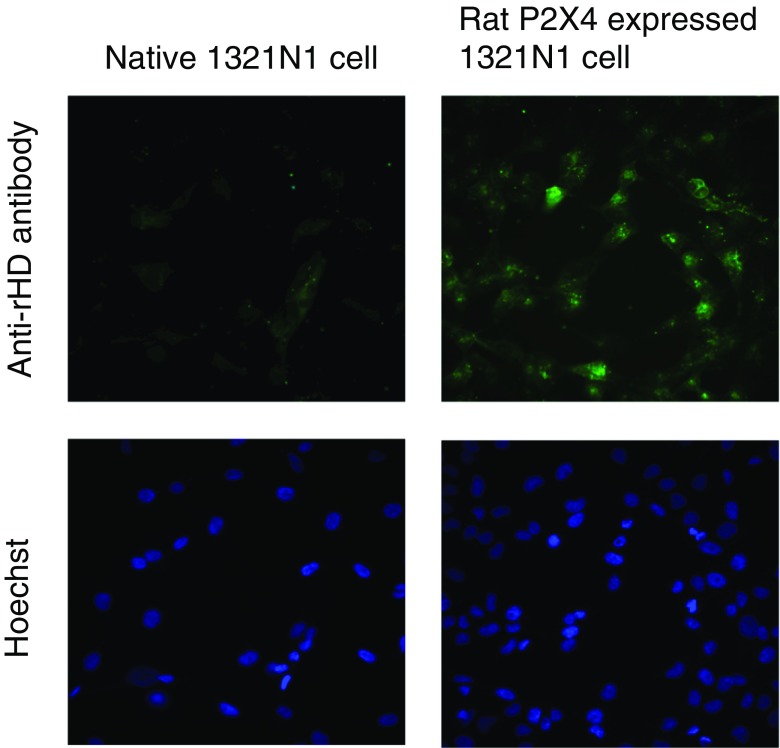
Fig. 5.
Detection of rP2X4 expressed on the cell by monoclonal antibody. Anti-rHD monoclonal antibody (12-10H, 10 μg/ml) and Alexa488-conjugated anti-mouse IgG were used for staining rP2X4 expressed on the 1321N1 cell (upper right). A native 1321N1 cell was also stained using both antibodies as a control (upper left). Hoechst was used to stain nuclei simultaneously (lower)

