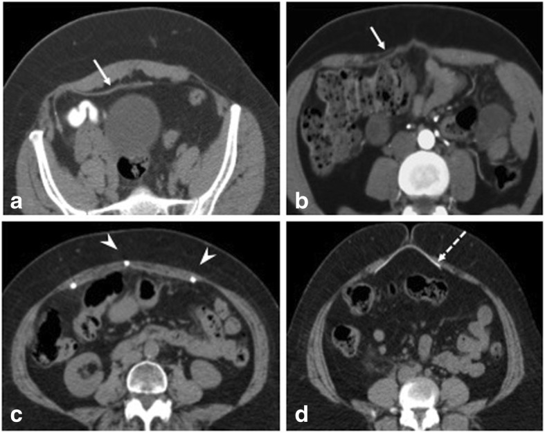
Fig. 3.

Mesh appearances on CT axial sections. a Preperitoneal placement of Prolene-based mesh appearing isodense to muscle. Presence of fat can aid in better visualization of the mesh. b Inlay Prolene-based mesh placement (arrow). c Laparoscopic intraperitoneal placement of mesh with tackers (arrowheads). d Mesh made of PTFE appearing radiodense (dashed arrow)
