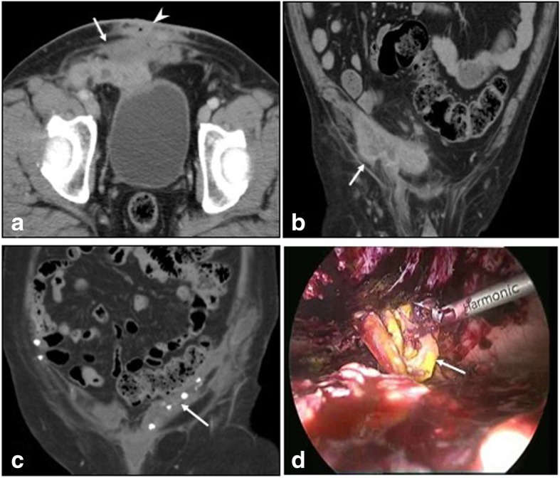
Fig. 9.

a, b CECT in axial and coronal sections show infected right inguinal hernia mesh repair with collections (arrows) and sinus tracks (arrowhead). Culture yielded Mycobacterium chelonae. c Coronal plain CT in another patient shows soft tissue thickening and collection along the mesh tackers (arrow). d Laparoscopic retrieval of infected mesh (arrow) which was covered with pus and granulation tissue
