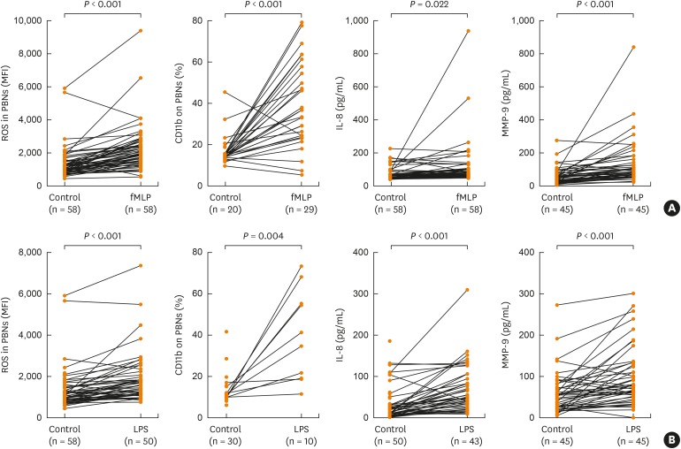
Fig. 1. In vitro PBN activation under fMLP or LPS stimulation. PBNs were isolated from asthmatics, stimulated under (A) fMLP stimulation or (B) LPS stimulation in 1 hour. The MFI of DCF fluorescence of PBNs and CD11b expression percentage were measured by flow cytometry; IL-8 and MMP-9 in the supernatants were measured by enzyme-linked immunosorbent assay. Data are presented as means ± standard deviation. P values were analyzed by paired t-test.
fMLP, N-formyl-methionyl-leucyl-phenylalanine; LPS, lipopolysaccharide; PBN, peripheral blood neutrophil; IL, interleukin; MMP, matrix metallopeptidase; ROS, reactive oxygen species.

