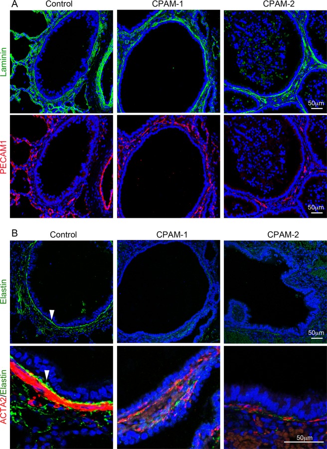
Figure 4.
Changes of extracellular matrix proteins in cystic airways of type 2 CPAM. (A) Laminin (green) was co-immunostained with vascular endothelial marker PECAM1 (red) and presented in separate panels. (B) Elastin (green) was co-immunostained with smooth muscle cell marker ACTA2 (red). Cell nuclei were counterstained with DAPI (blue). The lower panel shows a magnified small area from upper panel. Arrow: An elastin layer between epithelia and SMCs. Control samples were from normal age-matched lungs, and type 2 CPAM samples (CPAM-1 or -2) were from different patients.

