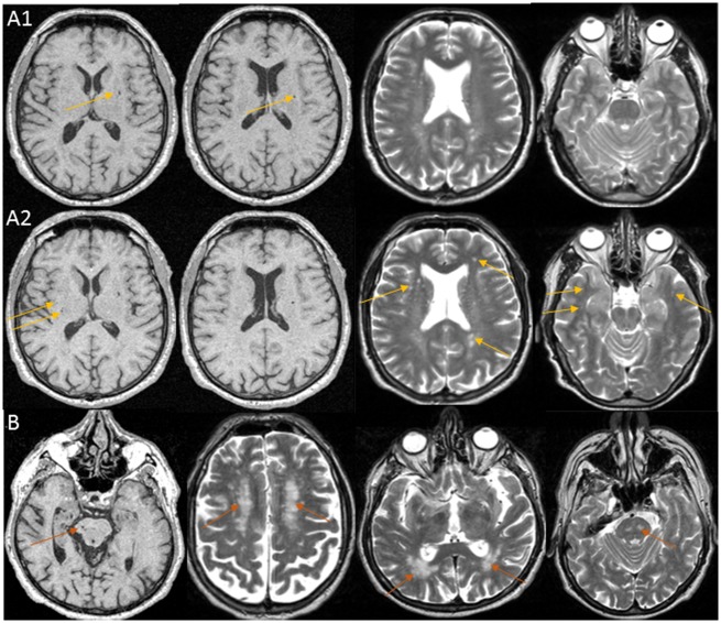
Figure 3.
MRI images of participants carrying heterozygote genotypes at CADASIL and CARASIL causing mutations. (A) Baseline (1) and 4-year follow-up (2) MRI scans of a 65-year old female participant with extensive SVD, in whom a NOTCH3 EGFr domain cysteine-modifying mutation was found: NM_000435.2 (NOTCH3):c.C2353T:p.R785C. Images show lacunar infarcts and dilated perivascular spaces in basal ganglia and white matter, and WMH in the periventricular region and deep white matter; on the follow-up MRI scan WMH and dilated perivascular spaces burden had increased and WMH became visible in the anterior temporal lobes (yellow arrows), a typical location for CADASIL. This participant remained free of stroke and dementia until the end of her follow-up at age 77. Her MMSE score was 28 at baseline and 26 at 12 years follow-up (secondary school education but no high school). (B) Baseline MRI scan of a 74-year-old female participant with extensive SVD, in whom a heterozygous CARASIL causing mutation was found: NM_002775.4 (HTRA1):c.1108C > T (p.Arg370Ter). Images show WMH and lacunes in the pons and extensive WMH in the deep white matter and periventricular region (magenta arrows). This participant was free of stroke and dementia at baseline but was lost to follow-up. Her baseline MMSE score was 27 (primary school education).

