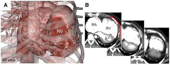
Figure 2.
Electrocardiogram-gated multi-detector computed tomography. (A) Three-dimensional imaging. Severely dilated right atrium and right ventricle are seen. (B) Of note, the localized coexistence of the residual muscular wall, severely thin right ventricle wall, and partial defects of the right ventricle muscular wall were also seen (red arrow). AP, anterior-posterior; LA, left atrium; LV, left ventricle; RA, right atrium; RV, right ventricle.

