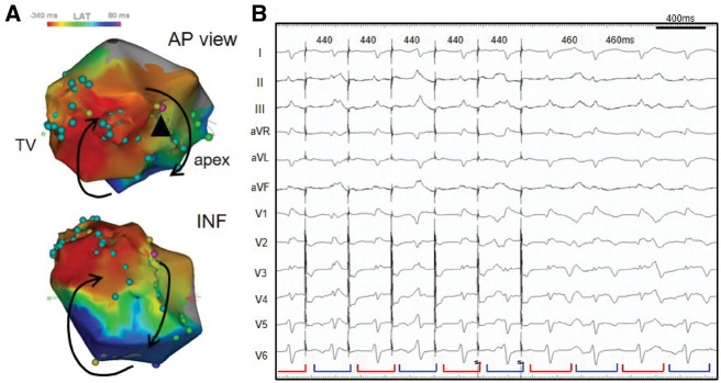
Figure 4.
(A) Electroanatomic activation map during the ventricular tachycardia. Counterclockwise activation on the inferolateral aspect of the right ventricle can be seen. (B) QRS alternans on the 12-lead electrocardiogram during entrainment pacing. Concealed entrainment was obtained by pacing from a low-voltage area in the anterior right ventricle (pink tag: arrowhead). The same QRS alternans as the ventricular tachycardia was reproduced by the pacing (red and blue brackets). AP, anterior-posterior; INF, inferior; LAT, local activation time; TV, tricuspid valve.

