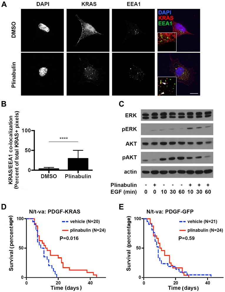
Figure 3.
Plinabulin yields endosomal accumulation of KRAS. (A) A549 cells were treated with 0.2% DMSO (upper panel) or 1.25 µM plinabulin (lower panel) for 2 h prior to immunofluorescence staining of DNA utilizing DAPI, (blue), EEA-1 (green) and KRAS (red). Scale bar, 10 µm. Inlays: 5x magnification; arrowheads indicate co-localization of EEA-1 and KRAS. (B) Quantification of the percentage EEA1/KRAS double-positive pixels of total KRAS-positive pixels in five high power fields (magnification, x100) from two independent experiments. (C) Immunoblot analysis of A549 cells serum-starved overnight and treated with 1% DMSO or 1.25 µM plinabulin for 2 h prior to treatment with 100 ng/ml EGF for indicated times. Mouse gliomas were generated in N/t-va;Ink4a/Arf-/- mice by transduction with RCAS-PDGF and (D) RCAS-KRASG12A, or (E) RCAS-GFP. Mice were treated with 5% dextrose solution i.p. (vehicle) or plinabulin 7.5 mg/kg i.p. for 5 subsequent days. Weight loss was utilized as an indicator of tumor formation to determine the time-point of treatment initialization.***P<0.0001. EGF, epithelial growth factor; KRAS, Kirsten rat sarcoma viral oncogene homolog; DAPI4, 6-Diamidino-2-phenylindole; ERK, extracellular signal regulated kinase; GFP, green fluorescent protein; PDGF, platelet derived growth factor; DMSO, dimethyl sulfoxide; EEA1, early endosomal antigen 1; pAKT, phosphorylated protein kinase B.

