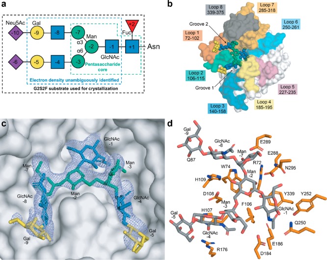
Figure 3.
Crystal structure of EndoS2 with complex biantennary glycan. (a) Structure of a full complex biantennary glycan, with annotations for the parts used for crystallization (black box), the parts unambiguously identified in the crystal structure (blue box), and the pentasaccharide core (green box). (b) Overall structure of the glycan bound within the active site crevasse of the GH domain. Annotation of GH domain loops for EndoS2 is colored. (c) Blue mesh illustrates the composite omit map of electron density (2Fo–Fc) contoured to 1σ and carved to 1.8 Å surrounding the CT glycan. (d) Key residues of EndoS2 interacting with CT product are colored in orange.

