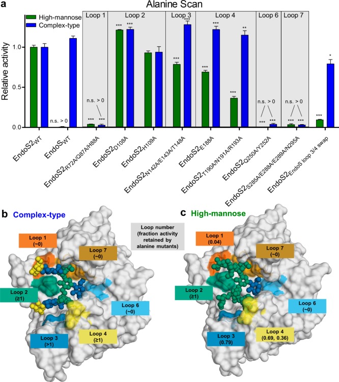
Figure 4.
Alanine scan mutagenesis of EndoS2 active site for complex-type and high-mannose IgG1. (a) Residues on each loop predicted to make contact with either glycan were mutated individually, or in batches, to alanine, and activity was measured using mass spectrometry, normalized to wild-type EndoS2. Statistical significance compared to wild-type EndoS2 is annotated (multiple comparisons test, Tukey method; *, p < 0.05; **, p < 0.01; ***, p < 0.001; n.s. > 0, not significantly greater than no-enzyme control). Mutated residues are colored by loop number, with fractional activity retained compared to wild-type EndoS2 in parentheses for (b) complex-type substrate and (c) high-mannose substrate.

