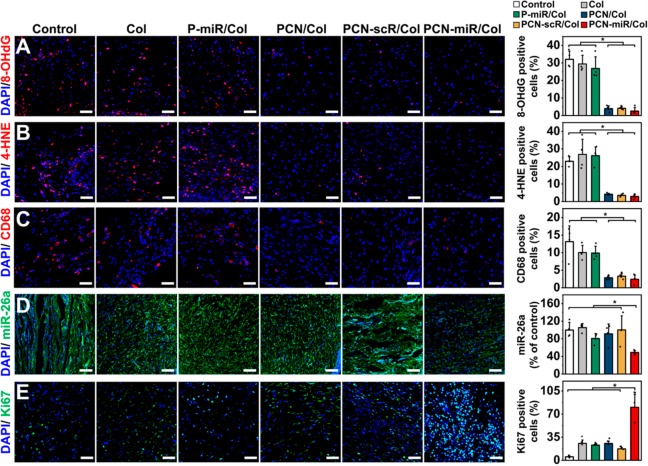
Figure 4.
In vivo topical application of PCN-miR/Col reshapes the highly oxidative and inflammatory wound microenvironment and corrects miR-26a overexpression. Representative confocal images of immunofluorescence staining and quantification for (A) 8-OHdG (a marker of oxidative DNA damage), (B) 4-HNE (a marker of lipid peroxidation), (C) CD68-positive macrophages, (D) miR-26a, and (E) Ki67-positive cells in sections from each group after 28 days of treatment (n = 4). Scale bars for 8-OHdG, 4-HNE, CD68, and Ki67 images, 50 μm; Scale bars for miR-26a image, 100 μm. All results are presented as mean ± SD, *P < 0.05 by two-tailed unpaired Student’s t tests.

