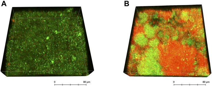
FIGURE 3.
3D-reconstruction of a LIVE/DEAD-stained confocal image stack of a supragingival oral biofilm after treatment with CHX. The biofilm was formed in situ on a bovine enamel slab for 72 h (methodology as described in Al-Ahmad et al., 2015) and was either left untreated (A) or was treated with 0.2% CHX for 5 min (B). Bacteria with intact (green; considered “live”) or compromised bacterial membranes (red; considered “dead”) are depicted indicating “pockets of viable cells” within the biofilm.

