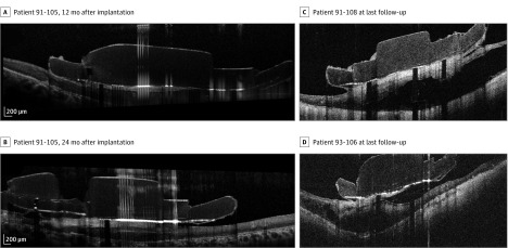
Figure 1. Fibrosis Evolution and Schisis at Follow-up.
For patient 91-105 on optical coherence tomography, fibrosis initially appeared as a thin hyperreflective line that thickened over time, becoming a real hyperreflective fibrotic plaque 12 months after implantation (A) and 24 months after implantation (B). C and D, Images of schisis at the last available follow-ups for patient 91-108 and patient 93-106.

