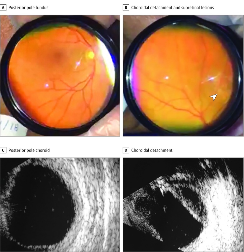
Figure 1. Findings in Patient 1.
A, Posterior pole fundus (inverted view). B, Midperipheral 360° choroidal detachment and yellowish subretinal lesions (arrowhead) (inverted view). C, Increased thickness of the posterior pole choroid (1.7 mm). D, Midperipheral choroidal detachment in the right eye and low-reflectivity echoes.

