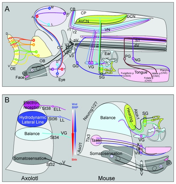
Figure 2. Distribution of sensory maps and the development of hindbrain sensory maps.
( A) Schematic presentation of the main features of the six cranial senses projected onto an embryonic mouse brain. (Pale yellow, left) Distributed olfactory sensory neurons of the olfactory epithelia coalesce their axons before reaching a specific olfactory glomerulus in the olfactory bulb (OB). (Pale lavender) Axons of retina ganglion neurons leave the eye orderly to project via the optic nerve to the optic chiasm (OC). Crossed contralateral axons form the orderly optic tract that distributes axons within the midbrain using matching gradients of several factors. (Gray) The trigeminal ganglion has three distinct branches and matching sensory neuron populations that reach different areas of the face. The central axons form in a temporal progression resulting in an inverted presentation of the face. (Pale pink) Taste buds of the tongue and pharynx are innervated by three cranial nerves that form a somewhat orotopic central projection to the solitary tract. (Light blue) The five vestibular sensory organs are innervated by somewhat orderly distributed sensory neurons that project via the vestibular nerve. Within the brain, vestibular afferents from different ear organs are partially segregated and partially overlapping in the various vestibular nuclei as well as the posterior lobes of the cerebellum. (Pale green) The organ of Corti of the cochlea is innervated by a temporally generated longitudinal array of spiral ganglion neurons that project in an orderly organization to dorso-ventral distinct regions of the cochlear nucleus complex, projecting a one-dimensional frequency array along the cochlea onto a matching frequency array of afferents in the cochlear nuclei. ( B) (Left) In the axolotl, there is a timing factor of afferent ingrowth such that the most ventral trigeminal projection reaches the hindbrain first (V at stage 32) whereas the most dorsal projection from the electroreceptive (lateral line) ampullary organs reaches the most dorsal part of the hindbrain last (ELL, stage 38). The inner ear vestibular ganglia (VG, stage 34) and mechanosensory lateral line ganglia (LL, stage 36) are reaching the alar plate between those extremes. (Right) In the mouse, the dorso-ventral patterning of the hindbrain is driven by countergradients of Wnt/BMP and Shh to regulate expression of transcription factors defining various nuclei. How these gradients define the positon of central nuclei and afferents is not completely clear. A temporal gradient of afferent development and projection development has thus far been demonstrated only for the spiral ganglion, taste and trigeminal system where the first neurons to form are the first to project to the most ventral part of their respective tract. Note that the auditory nuclei show an apparent inversion such that the most ventral projection from the basal spiral ganglion ends up in the more dorso-medial part of the cochlear nuclei because of the morphogenetic changes in cochlear nucleus neuron position. A, anterior; AC, anterior crista; Ascl1/Mash1, achaete-scute family basic helix-loop-helix transcription factor 1; Atoh7, atonal basic helix-loop-helix transcription factor 7; AVCN, antero-ventral cochlear nucleus; BMP, bone morphogenic protein; C, cochlea; CB, cerebellum; CN V, VII, IX, X, cranial nerve V, VII, IX, X; CP, choroid plexus of IV ventricle; D, dorsal; DCN, dorsal cochlear nucleus; dV, descending trigeminal tract; ELL, electroreceptive (ampullary organ) lateral line; GG, geniculate ganglion; HC, horizontal crista; L, lateral; LL, (mechanosensory) lateral line; M, medial; N, nasal; Neurog1/2, Neurogenin 1/2; NG, nodose ganglion; OB, olfactory bulb; OC, optic chiasm; OE, olfactory epithelium; P, posterior; PC, posterior crista; PG, petrosal ganglion; pV, principal trigeminal nucleus; r1, rhombomere 1; r2, rhombomere 2; S, saccule; SG, spiral ganglion; Shh, sonic hedgehog; ST, solitary tract; T, temporal; TIx3, T-cell leukemia homeobox 3; U, utricle; V, ventral; V1, ophthalmic branch of trigeminal nerve; V2, maxillary branch; V3, mandibular branch; VG, vestibular ganglion; VN, vestibular nucleus complex. Modified after 5, 12, 17, 33, 36, 54, 56, 63– 66.

