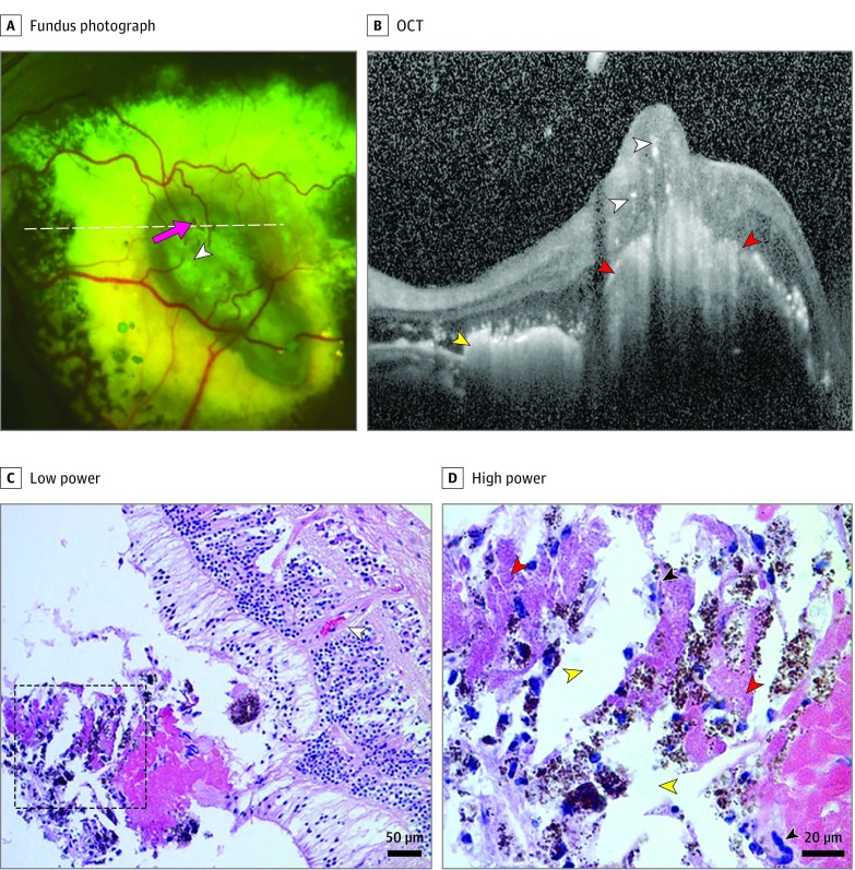
Figure 4. Fibrotic Nodules With Intraretinal Vessels in Coats Disease.
Fundus photograph (A) OCT (B) from a 7-year-old boy. Notable findings include retinal vessels diving into the lesion (white arrowhead), an adjacent dot of blood (pink arrow) on photograph, nodule (red arrowheads) with atrophy of overlying outer retinal layers, possible retinal vessels traveling at a right angle from the inner retina into the nodule (white arrowheads), and adjacent subretinal exudates (yellow arrowheads). Low-power (C) and high-power (D) light micrographs with a hematoxylin- eosin stain from an enucleated biorepository eye. A vessel traveling from the inner retina toward the nodule (white arrowhead) and multinucleated giant cells (black arrowheads) surrounding cholesterol clefts (yellow arrowheads) with a deposition of fibrinous material (red arrowheads) are observed on light micrographs. The horizontal white dotted line on the photograph corresponds to the OCT line scan while the black dotted box on the low-power micrograph (C) denotes the area shown in the high-power micrograph (D).

