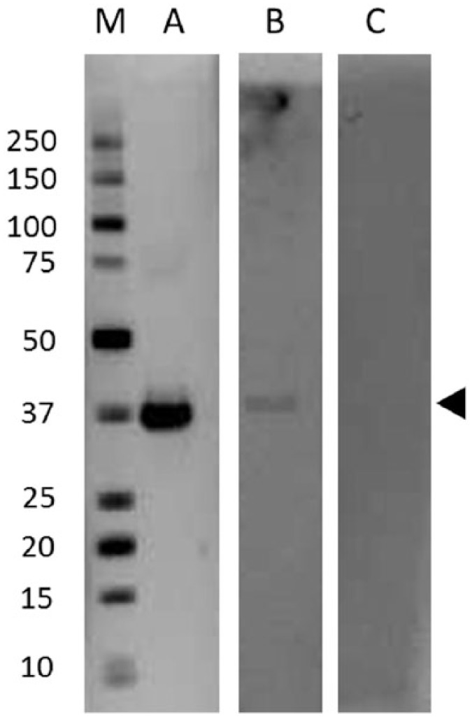
Figure 1.

Western blot indicating immunoreactivity to epsilon toxin from different sera. Lane A shows epsilon toxin (arrowed) reacted with a strongly positive serum (BUH00226), lane B shows epsilon toxin reacted with a weakly positive serum (BLT00139) and Lane C shows an example of a serum (BUH00239) which did not react with epsilon toxin. Molecular size markers (kDa) in Lane M.
