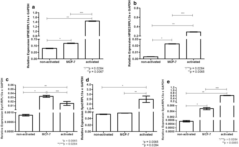
Fig. 6.
Relative expression of heparanases and syndecans in the exosomes. A pool of lymphocytes obtained from two healthy women donors was used to obtain non-activated lymphocytes or lymphocytes that were co-cultured with MCF-7 cells (activated lymphocytes). Exosomes were purified from conditioned medium of (non-activated T-lymphocytes); breast cancer cell line (MCF-7); or (activated T-lymphocytes). It is important to observe that T-lymphocytes activation was performed during 4 h at 37 °C and subsequently lymphocytes were plated in culture with DMEM medium, containing 10% fetal bovine serum and maintained overnight at 37 °C. The supernatant containing the cell-free media was used to obtain exosomes using Total Exosome Isolation kit. Quantitative RT-PCR was performed using total RNA extraction purified from exosomes preparation. a heparanase enzyme (HPSE); b heparanase-2 (HPSE2); c syndecan-1 (Syn1); d syndecan-2 (Syn2); e syndecan-4 (Syn4). The constitutive endogenous genes RPL13a and GAPDH were used to obtain the relative expression. The values represent average and standard deviation of triplicate assays. Statistical analysis was performed using Kruskal–Wallis test with Dunn auxiliary test

