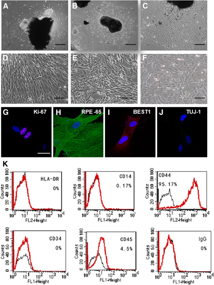
Fig. 1.
Isolation and characterization of cells from adult human RPE tissues. (a-c) Phase-contrast microscopy (A) of cultured RPE monolayer tissues. A small number of cells migrated out from the edge of the tissues. b, c While the cell layer expanded, the center tissues shrunk. The cell layer exhibited classical epithelial morphology with flat cell bodies possessing processes extending out from the cell body (scale bar: 100 μm). d-f Representative microscopy images of RPE cells at passages P1 (d), P10 (e), and P20 (f) (scale bar: 50 μm). g-j Characterization of cultured RPE cells by immunofluorescent staining using an antibody against Ki67 for proliferation (g), RPE65 (h) and BEST1 (i) for retinal pigmented epithelium, and Tuj1(j) for pan-neural cells (scale bar: 20 μm). k Flow cytometry analysis of the expression of HLA-DR, CD14 for endothelial, CD34 and CD45 for hematopoietic, and CD44 for proliferative progenitor cells

