Fig. 2.
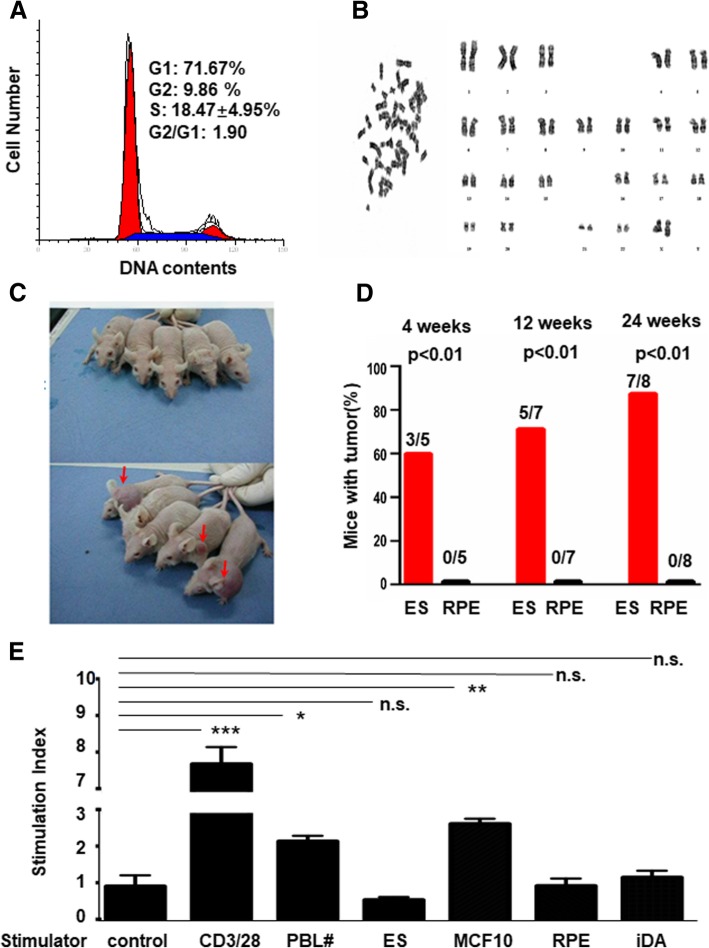
Evaluation of the safety and immunogenicity of cultured RPE cells. a Representative cell cycle progression of the P20 passage of RPE cells. b Representative G-banding analyses of karyotype for P20 passage RPE cells. c A picture of tumor formation in SCID-nude mice 4 weeks after injection of RPE (top panel) and ES cells (bottom panel) (n = 5 animals for each condition) Mice with tumors were arrowed (bottom panel). d Statistical analysis of the percentage of SCID-nude mice with tumors that formed from the ES and RPE cells measured at 4 (n = 5 animals for each condition), 12 (n = 7 animals for each condition), and 24 (n = 8 animals for each condition) weeks after injection. e Measurement of hPBL to co-cultured CD3/28, mismatched individual hPBL, ES, MCF10A, RPE, and iDA by one-way mixed lymphocyte reaction. The results were presented as a stimulation index. p values were determined using one-way ANOVA. Data are expressed as mean + SD; *p < 0.01, **p < 0.001, ***p < 0.0001 n.s., not significant; n = 3)

