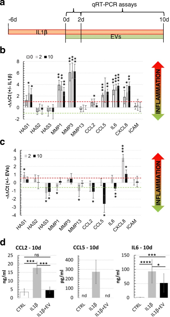
Fig. 7.

IL-1β and EV effect on FLSs. a Experimental plan for IL-1β inflammation and EVs supplementation in FLSs. IL-1β or IL-β + EVs (100,000 EVs:FLS) were freshly added with medium change each 48 h. b IL-1β at low concentration activates inflammation markers. Synoviocytes were treated with 25 pg/mL IL-1β and, after 6 days, at time points 0, 2, and 10 days, 11 genes related to inflammation were scored by qRT-PCR. The data are presented as −ΔΔCt relative to the untreated control for each time point. *p ≤ 0.05, **p < 0.01, ***p < 0.001. c EVs are able to reduce secretion of chemokines and cytokines under inflammation stimuli. FLSs treated with 25 pg/mL of IL-1β for 6 days were supplemented with EVs pooled from three ASC supernatants and OA-related genes scored after 2 and 10 days. Quantification of data is shown as mean ± SD and normalized for TBP. The data are presented as −ΔΔCt relative to IL-1β only treated FLSs. *p ≤ 0.05, **p < 0.01, ***p < 0.001. d ELISA assays confirm reduction of inflammation-related CCL2/CCL5 chemokines and IL-6 after 10-day EV exposure. Quantification of data is shown as mean ± SD. *p ≤ 0.05, ***p < 0.001, ****p < 0.0001
