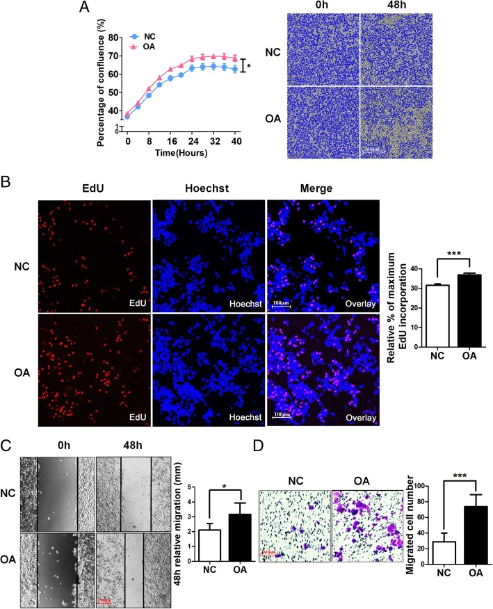
Fig. 2.
OA accelerates HeLa cell proliferation and migration. a Growth curves for HeLa cells treated with or without OA were analysed using an IncuCyte incubator microscope (the mean confluence values were compared at 48 h.; n = 5). b DNA synthesis of HeLa cells subjected to the EdU incorporation assay. EdU staining (red). Cell nuclei were stained with Hoechst33342 (blue). The quantification of EdU-positive cells was conducted macroscopically and was expressed as a percentage relative to the control cells. c OA promotes scratch-induced migration of HeLa cells. Representative phase contrast micrographs of cells treated with or without OA for 0 h. and 48 h.. Relative migration distances are calculated representing scratch closure after 48 h. compared with the initial distances. d Transwell assays were performed to investigate the effects of OA on the migration ability of HeLa cells. Quantitative analysis of migration experiments demonstrated that OA promotes HeLa cell migration compared with NC. *P < 0.05, ***P < 0.001

