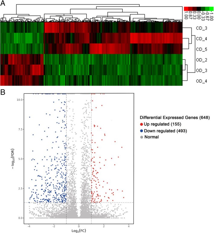
Fig. 4.
Heat map and volcano plots showed the expression profiles of mRNAs under two diet conditions. a Heat map for the DEGs. Each row represents a tissue sample (total 6 samples); each column represents a single gene. The gradual colour ranged from red to green represented the mRNA expression changing from up-regulation to down-regulation. b The volcano plot showed significantly changed mRNAs with FDR < 0.05 and |log2FC (fold change)| ≥ 1. The red-marked nodes represented up-regulated genes (155), the blue ones represented down-regulated genes (493), and the grey ones showed no significance

