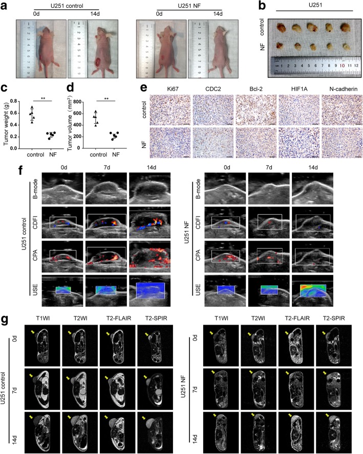
Fig. 7.
NF inhibited the growth of U251 xenograft tumors in nude mice. a-d U251 cells were subcutaneously injected into the mouse left flank to establish xenograft models. After NF treatments (15 mg/kg, once a day for a total of 2 weeks), all mice were observed, and the tumor weight and volume were measured and compared. **P < 0.01 vs. control group. e Expression of Ki67, CDC2, Bcl-2, HIF1A, and N-cadherin in xenograft tumors were analyzed by immunohistochemistry (original magnification: × 400, scale bar: 100 μm). f Ultrasonography evaluation included B-mode, CDFI, CPA, and USE. g T1WI, T2WI, T2-FLAIR, and T2-SPIR imaging of tumors by MRI

