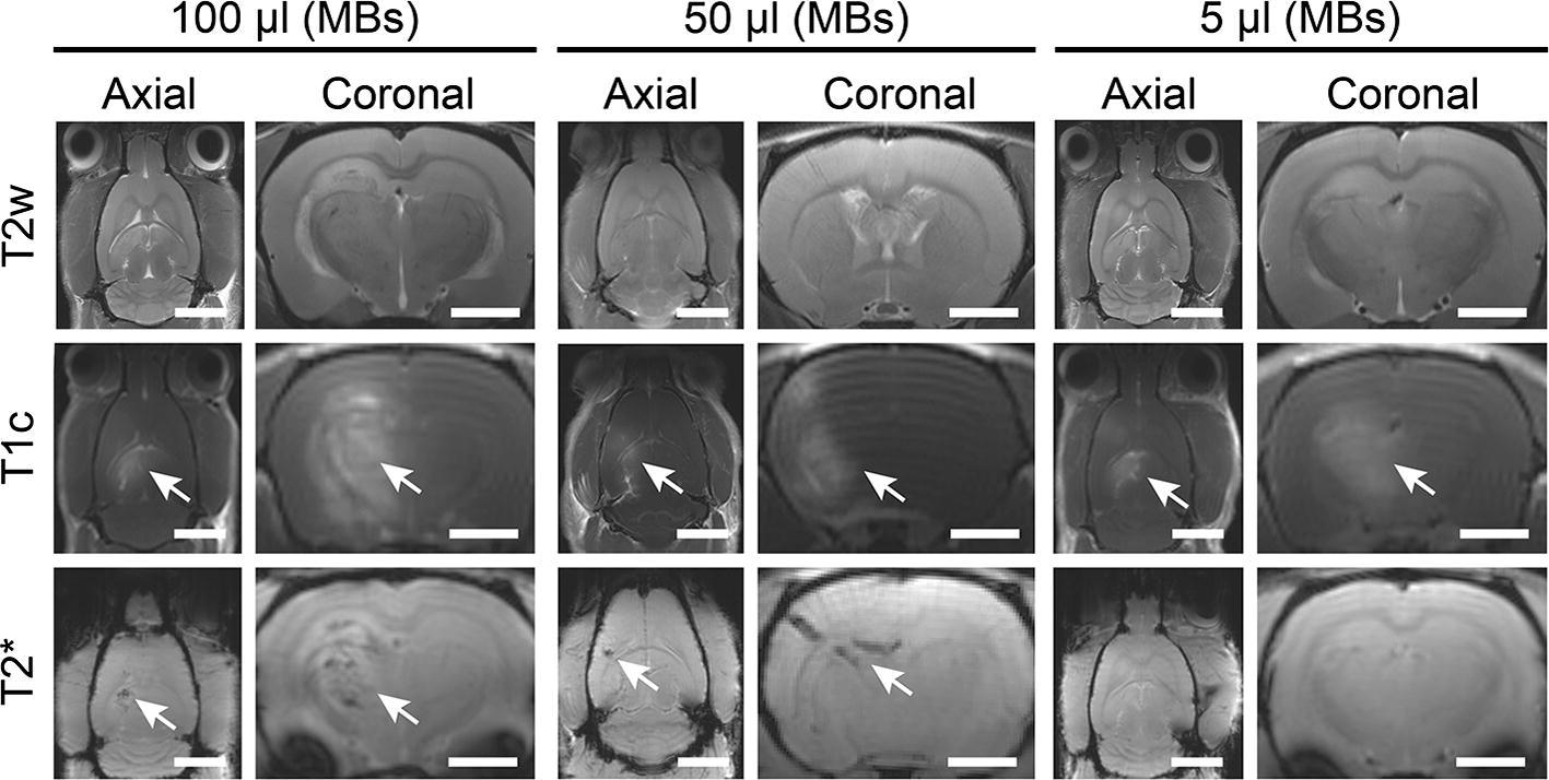
Fig. 9.

Representative axial and coronal MR images of animals treated for BBB opening at various MB doses: 100 µL, 50 µL and 5 µL. MR images reveal BBB opening (T1c hyperintensity) and microhemorrhage (T2* hypointensity). T2 images for inflammation were inconclusive. Scale bars for the axial and coronal images correspond to 5 mm and 2.5 mm, respectively
