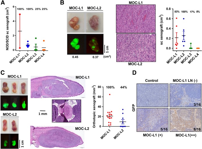
Fig. 3.
Xenotransplantation of cell lines. a Subcutaneous xenografic induction of cells admixed with Matrigel in NOD-SCID mice. Tumors are harvested 2 weeks after inoculation. * denotes that two recipients who show rapid tumor growth and die during the first two weeks; these are excluded for the analysis. The tumorigenicity of MOC-L1 and MOC-L2 is greater than that of MOC-L3 and MOC-L4. b Subcutaneous xenografic induction of cells in C57BL/6 mice. Left, gross pictures (Upper) and fluorescence images (Lower) of the resected tumors. Middle, histopathological pictures of the tumors. X200. Right, quantification. MOC-L1 and MOC-L2 exhibit syngeneic tumor induction, while MOC-L3 and MOC-L4 are nearly non-tumorigenic. c Orthotopic xenografic induction of MOC-L1 and MOC-L2 cells in C57BL/6 mice. Left, gross pictures (Upper) and fluorescence images (Lower) of the resected tongues. Middle, histopathological sections of the tongues revealing the presence of the tumors within each tongue. Note the presence of a positive neck node in a mouse implanted with MOC-L1 cells. Right, quantification of the tongue tumors. X100. d GFP immunohistochemistry of the neck lymph nodes of nude mice that had undergone orthotopic MOC-L1 xenotransplantation. -, negative, +, weak positive; ++, strong positive. Control, mice without xenotransplantation

