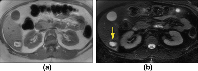
Figure 1.

Hepatic adenoma with haemorrhage in a 39-year-old woman. Axial T1WI in-phase (a) and T2WI with fat suppression (b) show a mass that is hyperintense with low signal in the periphery. These findings are consistent with bleeding within the lesion and rim of haemosiderin deposition.
