
Figure 3.
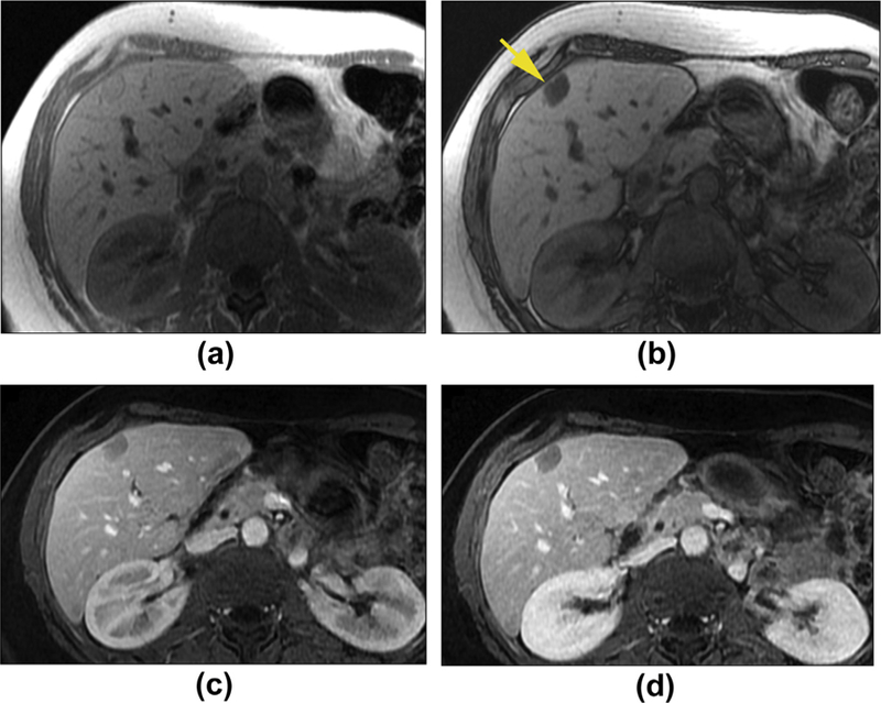
Hepatic adenoma with fat infiltration in a 49-year-old woman. Axial T1W in-phase (a) and out-phase (b) images show a mass with signal drop on out-phase image (arrow), which is consistent with a lipid-containing lesion. The capsule is noted as a rim of low signal intensity around the lesion on in-phase image (a). Axial enhanced T1WI in the hepatic late arterial phase (c) and delayed phase (d) shows the lesion to have minimal enhancement. Of note, the late arterial phase image acquisition might be the reason for the apparent lack of hypervascularity in this lesion.