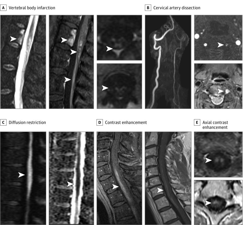
Figure 2. Confirmatory Magnetic Resonance Imaging Findings and Typical Gadolinium Enhancement Pattern in Spinal Cord Infarctions.
Confirmatory spinal cord infarction (SCI) findings are shown, including vertebral body infarction on short-τ inversion recovery imaging with associated gadolinium enhancement of the vertebral body infarct, SCI, and anterior cauda equina (A); cervical artery dissection with significantly decreased left vertebral flow with confirmed intramural hematoma shown on T1-fat suppression imaging (B) adjacent to SCI; and diffusion restriction on diffusion-weighted imaging with correlation on apparent diffusion coefficient (C). Spinal cord infarction gadolinium enhancement demonstrated with a typical craniocaudal linear strip on sagittal views (D) and corresponding anterior predominant gray matter and anteromedial spot (E) patterns on axial views, highlighting the predominant areas of ischemia.

