Figure 2.

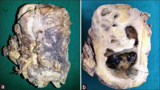
Gross photographs: (a) enlarged kidney with adherent capsule and (b) cut section shows dilated pelvicalyceal system with impacted stone, areas of fibrosis, and multiple yellow areas

Gross photographs: (a) enlarged kidney with adherent capsule and (b) cut section shows dilated pelvicalyceal system with impacted stone, areas of fibrosis, and multiple yellow areas