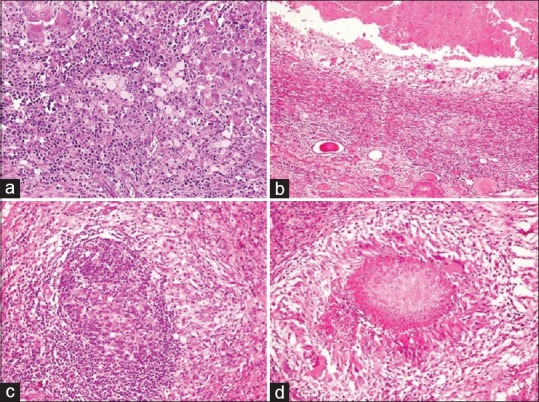
Figure 3.

Histopathology sections in xanthogranulomatous pyelonephritis: (a) renal parenchyma with plentiful xanthoma cells admixed with lymphoplasmacytic cells and giant cells (H and E, ×400), (b) abscess formation (H and E, ×200), (c) lymphoid follicle (H and E, ×400), and (d) granuloma formation (H and E, ×400)
