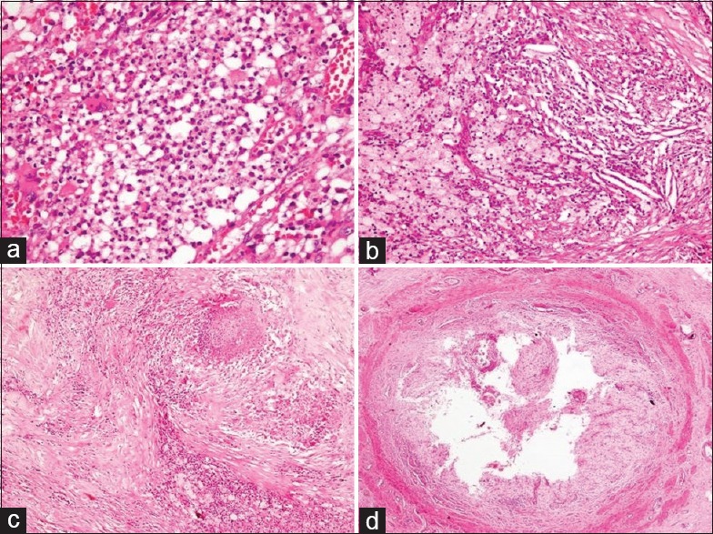
Figure 4.

Histopathology sections in xanthogranulomatous pyelonephritis: (a) neutrophilic collections (H and E, ×400), (b) cholesterol clefts, xanthoma cells, and mixed inflammation (H and E, ×200), (c) fibrosis, patchy areas of necrosis and histiocytes (H and E, ×100), and (d) severe ureteritis with complete replacement of urothelial lining by foam cells (H and E, ×40)
