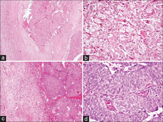
Figure 5.

(a and b) Xanthogranulomatous pyelonephritis with renal cell carcinoma: (a) tumor arranged in nests with adjoining renal parenchyma showing xanthogranulomatous pyelonephritis (H and E, ×40) and (b) tumor cell nests separated by thin septae (H and E, ×400), (c and d) xanthogranulomatous pyelonephritis with papillary urothelial carcinoma: (c) papillary tumor with adjacent xanthogranulomatous pyelonephritis (H and E, ×100) and (d) true papillae with fibrovascular cores and lined by layers of neoplastic urothelial cells (H and E, ×400)
